Anti-Cathepsin D
- Catalog #
- AB0043
- Category
- Antibodies
- Family
- Organelle Markers
- Source
- Goat
- Application
- WB, IHC (F), IHC (P), IF, IEM
- Reactivity
- Human, Rat, Mouse, Monkey, Canine
- Description
- Goat polyclonal antibody to Cathepsin D. Cathepsin D is a lysosomal aspartic protease of the pepsin family. Acid protease active in intracellular protein breakdown. It is composed of a dimer of disulphide-linked heavy and light chains, both produced from a single protein precursor. It is involved in the pathogenesis of several diseases such as breast cancer and possibly Alzheimer disease.
- Alternative names
- ceroid-lipofuscinosis, CLN10; CPSD, CTSD, lysosomal aspartyl protease, lysosomal aspartyl peptidase, neuronal 10 antibody.
- Gene Identifier/Accession#
- Gene ID: 1509, ENSG00000117984
- Form
- Polyclonal antibody supplied as a 200 or 500 µl (3 mg/ml) aliquot in PBS, 20% glycerol and 0.05% sodium azide. This antibody is epitope-affinity purified from goat antiserum.
- Concentration
- 3.00 mg/ml
- Isotype
- IgG
- Clonality
- Polyclonal
- Conjugation
- Unconjugated
- Immunogen
- Purified recombinant peptide derived from within residues 275 aa to the C-terminus of human Cathepsin D produced in E. coli.
- Antigen Sequence
- DQVEVASGLTLCKEGCEAIVDTGTSLMVGPVDEVRELQKAIGAVPLIQGEYMIPCEKVSTLPAITLKLGGKGYKLSPEDYTLKVSQAGKTLCLSGFMGMDIPPPSGPLWILGDVFIGRYYTVFDRDNNRVGFAEAARL
- Specificity
- This antibody gives a positive signal in the following human (Jurkat, HT1080, HUH, MDA-MB-231, ARPE19, SH-SY5Y), canine (MDCK) and monkey (COS-7) whole cell lysates.
- Storage
- For continuous use, store at 2-8 C for one-two days. For extended storage, store in -20 C freezer. Working dilution samples should be discarded if not used within 12 hours.
- Special instructions
- The antibody solution should be gently mixed before use. Avoid freeze/thaw cycles.
-
Application Values WB 1:250-1:1,000 IF 1:50-1:200 IHC (P) 1:200-1:1,000 IHC (F) 1:200-1:1,000 IEM 1:50-1:200 -
Sample WB IHC (F) IHC (P) IF ELISA IEM Human +++ +++ +++ +++ ND +++ Rat +++ +++ +++ +++ ND +++ Mouse +++ +++ +++ +++ ND +++ Canine +++ +++ +++ +++ ND +++ Monkey +++ +++ +++ +++ ND +++ -

Immunofluorescence – anti-CTSD Ab at 1/100 dilution in RAW264.7 cells; cells were fixed with PFA and permeabilized with 0.05% saponin; 
Anti-CTSD Ab at 1/500 dilution; endogenous CTSD (50 µg per lane); rabbit polyclonal to goat IgG (HRP) at 1/10,000 dilution; 
Immunogold labeling using melanocytes and anti-CTSD Ab; 
IHC of mouse spleen using anti-CTSD antibody and FFPE tissue after heat-induced antigen retrieval. Anti-CTSD Ab at 1:500/DAB detection; 
Immunofluorescence – anti-CTSD Ab at 1/100 dilution in NHI/3T3 cells; cells were fixed with methanol and permeabilized with 0.1% saponin; 
IHC of mouse lung using anti-CTSD antibody and FFPE tissue after heat-induced antigen retrieval. Anti-CTSD Ab at 1:500/DAB detection; 
Immunoperoxidase of polyclonal antibody to CTSD (1/200) on paraformaldehyde-fixed paraffin-embedded mouse lung; 
Anti-CTSD Ab at 1/500 dilution; endogenous CTSD (100 µg per lane) and transfected 293FT cell lysate (at 30 µg per lane); rabbit polyclonal to goat IgG (HRP) at 1/10,000 dilution; 
IHC of mouse brain using anti-CTSD antibody and FFPE tissue after heat-induced antigen retrieval. Anti-CTSD Ab at 1:500/DAB detection; 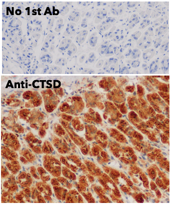
IHC of mouse stomach using anti-CTSD antibody and FFPE tissue after heat-induced antigen retrieval. Anti-CTSD Ab at 1:500/DAB detection; 
IHC of mouse eye using anti-CTSD antibody and FFPE tissue after heat-induced antigen retrieval. Anti-CTSD Ab at 1:500/DAB detection;
Usage
Reactivity Chart
Images
375 €
Quantity / Unit Size
600 µg
1.5 mg
Add to shopping cart
Shipping
No Shipping