Anti-CD63
- Catalog #
- AB0047
- Category
- Antibodies
- Family
- CD Markers
- Source
- Goat
- Application
- WB, IHC (F), IHC (P), IF
- Reactivity
- Human, Rat, Mouse, Monkey, Canine
- Description
- CD63 is a member of the transmembrane 4 superfamily, also known as the tetraspanin family. Most of the members are cell-surface proteins that are characterized by the presence of four hydrophobic domains. These proteins mediate signal transduction events that play a role in the regulation of cell development, activation, growth and motility. This protein is a cell surface glycoprotein that is known to complex with integrins. It may function as a blood platelet activation marker.
- Alternative names
- CD63 antigen, CD63 antigen (melanoma 1 antigen), granulophysin, LAMP-3, lysosomal-associated membrane protein 3, lysosome-associated membrane glycoprotein 3, ME491, melanoma-associated antigen, melanoma 1 antigen, melanoma-associated antigen ME491, MLA1, ocular melanoma-associated antigen, OMA81H, tetraspanin-30, TSPAN30 antibody.
- Gene Identifier/Accession#
- Gene ID: 967, ENSG00000135404
- Form
- Polyclonal antibody supplied as a 200 or 500 µl (3 mg/ml) aliquot in PBS, 20% glycerol and 0.05% sodium azide. This antibody is epitope-affinity purified from goat antiserum.
- Concentration
- 3.00 mg/ml
- Isotype
- IgG
- Clonality
- Polyclonal
- Conjugation
- Unconjugated
- Immunogen
- Purified recombinant peptide derived from within residues 120 aa to 175 aa of human CD63 produced in E. coli.
- Antigen Sequence
- MENYPKNNHTASILDRMQADFKCCGAANYTDWEKIPSMSKNRVPDSCCI
- Specificity
- Reacts with CD63, a 40-60 kDa glycoprotein, detected by Western blot in the following human (HeLa, HUH, Jurkat), mouse (AtT-20, Hepa, 3T3, RAW264.7), canine (MDCK) and monkey (COS-7) whole cell lysates.
- Storage
- For continuous use, store at 2-8 C for one-two days. For extended storage, store in -20 C freezer. Working dilution samples should be discarded if not used within 12 hours.
- Special instructions
- The antibody solution should be gently mixed before use.
-
Application Values WB 1:500-1:5,000 IF 1:25-1:250 IHC (F) 1:250-1:1,000 IHC (P) 1:250-1:1,000 -
Sample WB IHC (F) IHC (P) IF ELISA Human +++ +++ +++ +++ ND Rat +++ +++ +++ +++ ND Mouse +++ +++ +++ +++ ND Canine +++ +++ +++ +++ ND Monkey +++ +++ +++ +++ ND -
-

Anti-CD63 Ab at 1/2,500 dilution; lysate at 50 µg per lane; Rabbit polyclonal to goat IgG (HRP) at 1/10,000 dilution; 
IHC of mouse spleen using anti-CD63 antibody and FFPE tissue after heat-induced antigen retrieval. Anti-CD63 Ab at 1:500/DAB detection; 
Immunofluorescence – anti-CD63 Ab in Hepa1-6 cells at 1/50 dilution; cells were fixed with 4% of PFA; 
IHC of human pancreas using anti-CD63 antibody and FFPE tissue after heat-induced antigen retrieval. Anti-CD63 Ab at 1:500/DAB detection; 
Anti-CD63 Ab at 1/2,500 dilution; lysate at 50 µg per lane; Rabbit polyclonal to goat IgG (HRP) at 1/10,000 dilution; 
IHC of human cervix using anti-CD63 antibody and FFPE tissue after heat-induced antigen retrieval. Anti-CD63 Ab at 1:500/DAB detection; 
Immunofluorescence – anti-CD63 Ab in Hepa1-6 cells at 1/50 dilution; cells were fixed with 4% of PFA; 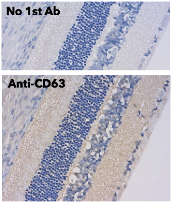
IHC of rat eye using anti-CD63 antibody and FFPE tissue after heat-induced antigen retrieval. Anti-CD63 Ab at 1:500/DAB detection;
Usage
Reactivity Chart
External References (18)
Images
375 €
Quantity / Unit Size
600 µg
1.5 mg
Add to shopping cart
Shipping
No Shipping