Anti-Bluebonnet
- Catalog #
- AB9139
- Category
- Antibodies
- Family
- Fluorescent Proteins
- Source
- Goat
- Application
- WB, IHC (F), IHC (P), IF, IEM
- Reactivity
- Transfected cells
- Description
- Goat polyclonal antibody to Bluebonnet. Bluebonnet is a monomeric blue fluorescent protein engineered from coral-derived fluorescent proteins originating from the cnidarian Entacmaea quadricolor. It is designed as a bright and photostable reporter suitable for fusion tagging, live-cell imaging, and multicolor experiments. Bluebonnet is a basic (constitutively fluorescent) protein with rapid maturation and stable fluorescence. It has a maximum excitation at 399 nm and a maximum emission at 454 nm. Bluebonnet-tagged proteins can be detected in Western blot at approximately 26–27 kDa plus the molecular weight of the fused protein. In immunofluorescence, antibody staining can enhance detection, particularly in fixed samples or low-expression systems. These antibodies are widely used in multicolor imaging, protein localization, and live-cell studies in mammalian and other model systems.
- Alternative names
- Bluebonnet2 antibody
- Gene Identifier/Accession#
- N/A
- Form
- Polyclonal antibody supplied as a 100 µl (3 mg/ml) aliquot in PBS, 20% glycerol and 0.05% sodium azide. This antibody is epitope-affinity purified from goat antiserum.
- Concentration
- 3.00 mg/ml
- Isotype
- IgG
- Clonality
- Polyclonal
- Conjugation
- Unconjugated
- Immunogen
- Affinity purified recombinant fluorescent protein produced in E. coli.
- Antigen Sequence
- N/A
- Specificity
- In lysates of transfected cells with the plasmid containing the fluorescent sequence, detects the recombinant protein by Western blot.
- Storage
- Store at -20 C for long-term storage. Store at 2-8 C for up to one month.
- Special instructions
- Avoid freeze/thaw cycles.
-
Application Values WB 1:500-1:5,000 IHC (F) 1:50-1:500 IHC (P) 1:50-1:500 IF 1:50-1:500 IEM 1:50-1:500 -
Sample WB IHC (F) IHC (P) IF IEM Transfected cells +++ +++ +++ +++ +++ -
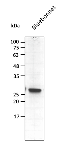
Anti-Bluebonnet Ab at 1/2,500 dilution using HEK293 transfected cell lysates at 40 µg per lane; rabbit polyclonal to goat IgG (HRP) at 1/10,000 dilution; 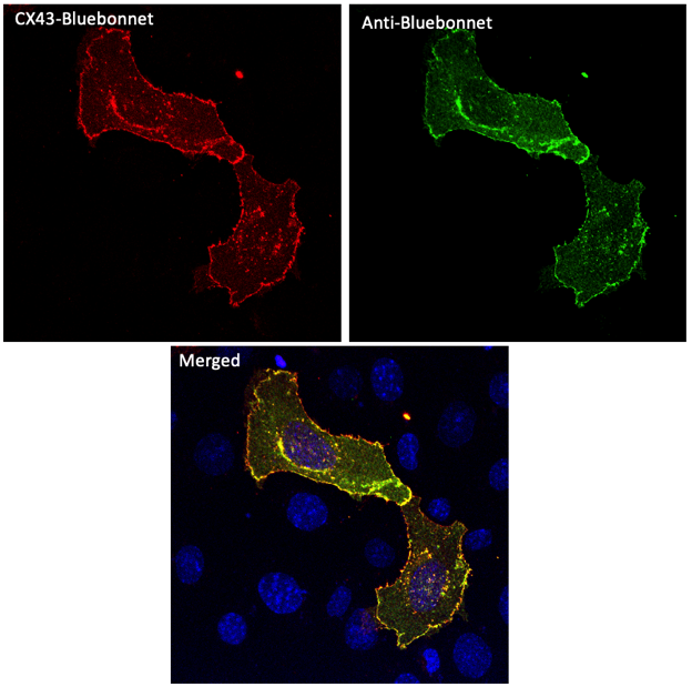
Immunofluorescence – anti-Bluebonnet Ab using hCEC cells transfected with CX43-Bluebonnet; cells were fixed with methanol and anti-Bluebonnet at 1/250;CX43-Bluebonnet (Ex 405 nm / Em 450–480 nm.) converted into red color;
Usage
Reactivity Chart
Images
375 €
Quantity / Unit Size
300 µg
Add to shopping cart
Shipping
No Shipping