Anti-CANX
- Catalog #
- AB0041
- Category
- Antibodies
- Family
- Loading Controls
- Source
- Goat
- Application
- WB, IHC (F), IHC (P), IF
- Reactivity
- Human, Rat, Mouse, Monkey, Canine
- Description
- Goat polyclonal to CANX (Calnexin) - endoplasmic reticulum (ER) membrane marker. CANX is a member of the calnexin family of molecular chaperones. This protein is a calcium-binding, ER-associated protein that interacts transiently with newly synthesized N-linked glycoproteins, facilitating protein folding and assembly. It may also play a central role in the quality control of protein folding by retaining incorrectly folded protein subunits within the ER for degradation.
- Alternative names
- Calnexin, CALX, CNX, FLJ26570, histocompatibility complex class I antigen binding protein p88, IP90, major histocompatibility complex class I antigen-binding protein p88, MS952, P90 antibody.
- Gene Identifier/Accession#
- Gene ID: 821, ENSG00000127022
- Form
- Polyclonal antibody supplied as a 200 or 500 µl (2 mg/ml) aliquot in PBS, 20% glycerol and 0.05% sodium azide. This antibody is epitope-affinity purified from goat antiserum and it is identical to AB4100-200.
- Concentration
- 2.00 mg/ml
- Isotype
- IgG
- Clonality
- Polyclonal
- Conjugation
- Unconjugated
- Immunogen
- Purified recombinant peptide within residues 550 aa to the C-terminus of human CANX produced in E. coli.
- Antigen Sequence
- KSDAEEDGGTVSQEEEDRKPKAEEDEILNRSPRNRKPRRE
- Specificity
- Detects a band of 90 kDa by Western blot in the following human (293A, primary fibroblasts, HaCat, HeLa, HMEC-1, Jurkat, MNT1, U-118, rat (TR-iBRB), mouse (3T3, AtT-20, Hepa, Raw264.7), monkey (COS-7) and canine (D17) whole cell lysates.
- Storage
- For continuous use, store at 2-8 C for one-two days. For extended storage, store in -20 C freezer. Working dilution samples should be discarded if not used within 12 hours.
- Special instructions
- The antibody solution should be gently mixed before use.
-
Application Values WB 1:500-1:5,000 IF 1:50-1:500 IHC (P) 1:200-1:1,000 IHC (F) 1:200-1:1,000 -
Sample WB IHC (F) IHC (P) IF ELISA Human +++ +++ +++ +++ ND Rat +++ +++ +++ +++ ND Mouse +++ +++ +++ +++ ND Canine +++ +++ +++ +++ ND Monkey +++ +++ +++ +++ ND -
-
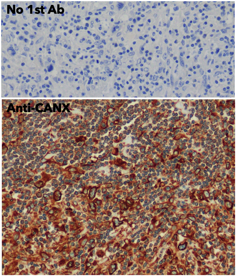
IHC of human lymph node using anti-CANX antibody and FFPE tissue after heat-induced antigen retrieval. Anti-CANX Ab at 1:750/DAB detection; 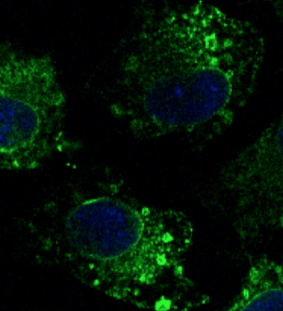
Immunofluorescence – anti-CANX Ab in Hepa1-6 cells at 1/100 dilution; cells were fixed with 4% of PFA; 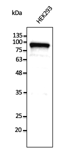
Anti-CANX Ab at 1/2,500 dilution; lysates at 50 µg per lane; rabbit polyclonal to goat IgG (HRP) at 1/10,000 dilution; 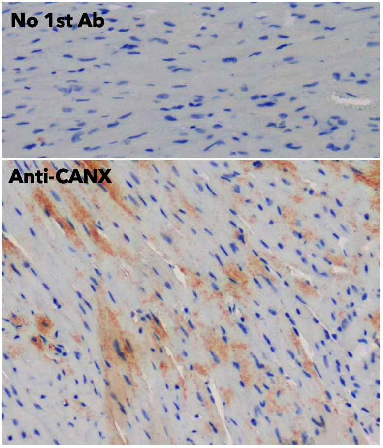
IHC of human heart using anti-CANX antibody and FFPE tissue after heat-induced antigen retrieval. Anti-CANX Ab at 1:750/DAB detection; 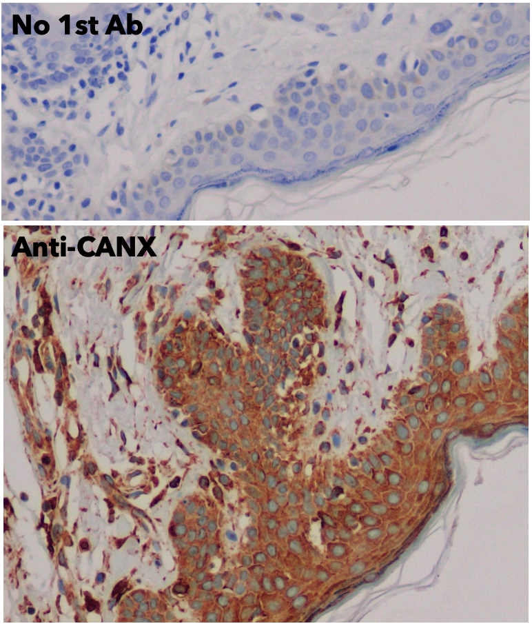
IHC of human skin using anti-CANX antibody and FFPE tissue after heat-induced antigen retrieval. Anti-CANX Ab at 1:750/DAB detection; 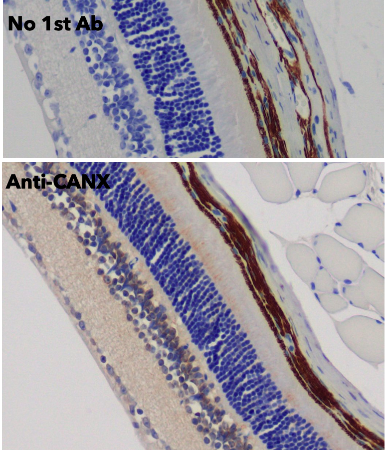
IHC of mouse retina using anti-CANX antibody and FFPE tissue after heat-induced antigen retrieval. Anti-CANX Ab at 1:750/DAB detection; 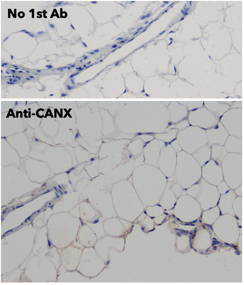
IHC of human adipose tissue anti-CANX antibody and FFPE tissue after heat-induced antigen retrieval. Anti-CANX Ab at 1:750/DAB detection; 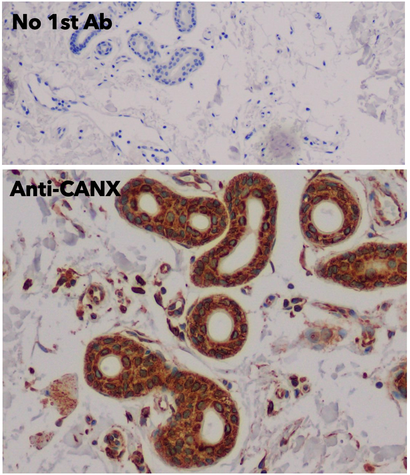
IHC of human adipose tissue anti-CANX antibody and FFPE tissue after heat-induced antigen retrieval. Anti-CANX Ab at 1:750/DAB detection;
Usage
Reactivity Chart
External References (38)
Images
375 €
Quantity / Unit Size
400 µg
1 mg
Add to shopping cart
Shipping
No Shipping